All type of modern facilities for eye care available :
REFRACTIVE SERVICES
Want to Get rid Of Spectacles?
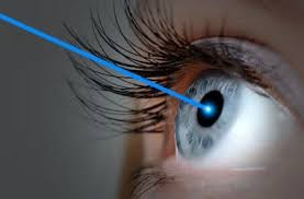
LASIK LASER
LASIK (Laser-Assisted In Situ Keratomileusis)
The Science Of Refractive Surgeries
. Reduces dependence on contact lenses or spectacles
.appropriate preoperative evaluation to determine the best technique
. Greek words for “cornea” (kerato) and “to carve” (mileusis).
. combines lamellar corneal surgery with the accuracy of the excimer laser.
. involves excimer laser ablation of cornea stroma beneath a hinged corneal flap that is
created with a microkeratome or a femtosecond laser
. most frequently performed keratorefractive procedure
. because of its safety, efficacy, quick recovery of vision, and minimal
patient discomfort.
. Excited dimer of argon and fluoride – releases UV energy at 193nm for
corneal ablation
. The 193nm wevelength – optimal for surgical use
. MOA- Non thermal Ablative Photodecomposition
PATIENT SELECTION
. >18 Years
. Stable Refractive status for at least 1 Year.
. Current FDA approval-
- Myopia upto -12D
- Hyperopia upto +6D
- Astigmatism upto 6D
CONTRAINDICATIONS
. Systemic factors-
– poorly controlled IDDM
-Pregnancy/ lactation
-Autoimmune/ connective tissue disorders (RA, SLE, PAN, etc.)
-Keloid tendency
-Systemic Infection (HIV, TB)
-Drugs-Azathioprene/ Steroids (slow wound healing)
.Ocular Factors-
– Glaucoma, RD (Suction Pressure-ON damage, Blebs)
-Previous h/o RD
-One eyed
-h/o Herpetic Keratitis (one year prior to surgery)
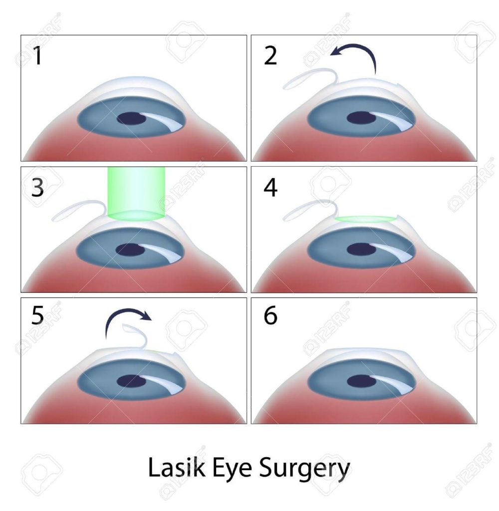
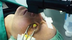
OPERATIVE TECHNIQUE
- Suction ring
- Flap Creation
- Excimer laser ablation
- Flap Repositioning
BASIC STEPS AND MACHINE SPECIFICATION
. Topical anaesthesia
.Surgical Painting and draping
.Lid speculum with aspiration
APPLICATION OF PNEUMATIC SUCTION RING
.lid speculum is depressed to proptose the eye to allow better
apposition of the ring to the globe
.suction ring is then placed on the eye
.patient is warned of a slight feeling of pressure and discomfort.
. At this point, four helpful sings assure the surgeon that adequate
suction has been obtained:
- The suction ring can be lifted toward the ceiling slightly and the eye should come up with it.
- The pupil will dilate slightly.
- The patient will confirm that everything has gone dark.
- The eye will feel firm to palpation.
CHECKING FOR ADEQUATE INTRAOCULAR PRESSURE
. Raise the IOP to 65mm Hg which is necessary for the microkeratome to create a pass and resect the corneal flap.
. This is crosschecked with Barraquers tonometer.
INSERTION OF THE MICROKERATOME HEAD AND CREATION OF THE KERATECTOMY
. Adequate lubrication
.Resection of cornea flap
.Several different microkeratomes are available
- Microkeratome
- Femtosecond Laser (Intralase)
MICROKERATOME
– Uses Disposable blades
–If using a two-piece microkeratome, the head is slid onto the the suction ring and advanced unit the gear on the microkeratome head engages the track.
–The surgeon then activates the microkeratome using forward and reverse foot control, the suction is turned off after the mocrokeratome pass, and then suction ring can be carefully removed
FEMTOSECOND LASER FOR FLAP
. Uses laser pulses to cause microcavitation bubbles at a preset depth in the corneal stroma
.The cavitation bubble is composed primarily of water and carbon dioxide.
.Creates photodisruption using femtosecond solid state with wavelength of 1053nm
. Needs lower vacuum.
.Very short pulse with spot size of 3
.High precision cutting device.
.Better LASIK outcomes
FEMTOSECOND LASER ADVANTAGES
. Reduced patient anxiety.
. Deep-set or small eyes, blepharospasm, or steep or flat corneas
. more control over flap centration, size, hinge location, and hinge width
LIFTING OF THE CORNEAL FLAP AND PREPARATION OF THE STROMAL BED
.Flap is reflected back with either a blunt, fine forceps or a cyclodialysis spatula
.Some surgeons reflect the flap back so that the stromal surfaces are opposed and the epitheial surface is exposed in a ‘taco’ fashion
LASIK FOR MYOPIA
.The normal cornea has a prolate shape (greater curvature centrally than peripherally)
.LASIK for myopia reverse this prolate cornea and decrease the central corneal curvature to create an oblate shape
LASIK FOR HYPERMETROPIA
. More than 4-5D-major challenge to the refractive surgeon.
. Consider the age of the patient in surgery for hyperopia
.must consider manifest, latent, and facultative hyperopia in planning surgery and to anticipate the long-term efficacy of the procedure
.it is common for a young patient to have latent to have hyperopia of 3.0 to 4.0 diopters exposed by cycloplegia.
. The amount of correction that should be provided during surgery is controversial in such cases.
HOW IS LASIK PROCEDURE DONE?
-
NO INJECTION NO PAIN
Just anaesthetic eye drops are sufficient to numb the eyes to undergo the procedure easily
CATARACT SERVICES
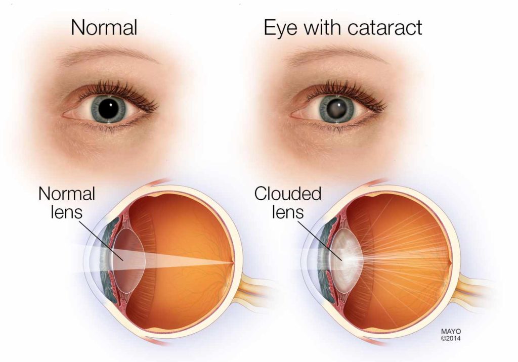
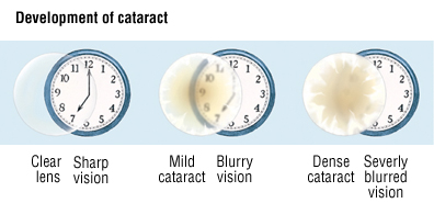
WHAT IS CATARACT ?
Cataract is defined as opacification of lens fibres.
SYMPTOMS OF CATARACT :
- Cloudy, blurred or dim vision
- Increasing difficulty with vision at night
- Increased sensitivity to light.
- Glare
- Need for brighter light for reading and other activities
- Seeing “halos” around lights
- Frequent changes in eyeglass or contact lens prescription
- Fading or yellowing of colors
- Double vision in a single eye
RISK FOCTORS
- Aging
- Associated ocular conditions
- Myopia (it is when the eye is not able to focus properly on obects in distance)
- Retinal detachment (the retina separates from the layer underneath)
- Infection
Toxic Factors
- Aspirin
- Corticosteroids
- Cigarette smoking
- Chemical burns
- Lonizing radiation
Nutritional factors
. Poor nutrition
.Physical Factors
- . Blunt trauma
- . Ultraviolet radiation in sunlight and x-ray
Systemic Disease and syndromes
- Diabetes
- Renal disorders
- Musculoskeletal disorders
Types
CONGENITAL CATARACT:
>33% Of the cases it’s idiopathic
>Hereditary causes: Chomosomal disorder (trisomy 21), sticker syndrom, lowe’s syndrome
>Maternal Factors (DRIM): Drugs like corticosteroids, Infection such as rubella,toxoplasmosis,Radiation,Malnutritionetc.may lead to congenital cataract
TYPE OF SENILE CATARACT
1. Cortical Senile cataracts
>It’s characterized by white, wedge-like opacities that start periphery of the lens and work their way to the center in a spoke-like fashion.They progress slowly and often do not cause severe loss of vision.
TYPE OF SENILE CATARACT
Posterior sub-capsular Cataract
>Posterior sub-capsular is cloudy area at the back of the lens capsule and cause visual
CLINICAL MANIFESTATIONS.
.Painless,blurry vision
. The person experiences reduced contrast sensitivity,and reduced visual acuity
.Astigmatism (it occurs when the cornea is irregularly shaped or sometimes because of the curvature of the lens inside the eye)
. Monocular diplopia (double vision in only one eye )
DIAGNOSTIC TESTS
.Snellen visual acuity test
.Opthalmoscopy
. Slit lamp examination
COMPLICATION OF CATARACT SURGERY
Immediate Preoperative
.Retrobulbar hemorrhage can result from retrobulbar infiltration of anesthetic agents.It can manifests as increased IOL, Proptosis,lid tightness and subconjunctival hemorrhageb with or without edema.
Intraoperative
. Rupture of the posterior capsule
Early postoperative
.Acute bacterial endophthalmitis characterized by marked visual loss, pain, lid edema.
Late Postoperative
. Suture related problems
.Malposition of the IOL
.Chronic endophthalmitis
Primary vs Secondary implantation
.Primary implantation – use of IOLs during surgery for cataract
.Secondary implantation – implantation of IOL to corect aphakia in a previosly operated eye
Parts of IOL
.OPTIC
Part of the lens that focuses light on the retina.
.HAPTIC
Small filaments connected to the optic that hold the lens in place in the eye
Silicone
. Elastomer Polydimetysiloxane
.Capable of large and reversible deformation
. Good memory
. Surface deposits are commin
. Addiotives in silicone IOL are
- – UV chromophore
- – Phenl group to increase Refractive Index from 1.41 to 1.46
Hydrogels
- HEMA or 2 Hydroxyethylmethacrylate
- Polymerization in ethylene glycon medium
- Hydrate to from soft, swollen, rubbery mass
- Hydrophilic hence repel cells and microbes
- Refactive Index -1.43 to 1.48
Truedge
- Truedge 360 Square Edge acts as a mechanical barrier for cell proliferation
- 10 Angulation, steep vault of 0.4 mm
Aurofold lens
- negative aspheric hydrophilic acrylic foldable intraocular lens made of p-HEMA material
- Truedge technology to prevent PCO
- zero Degree Angulation
Aurofold lens
- p-HEMA material
- Zero degree angulation
- Disposable delivery system
Auro toric
- Auroflex Toric IOL has been carefully designed with toricity on the anterior surface of the IOL.
- UV absorbing p-HEMA
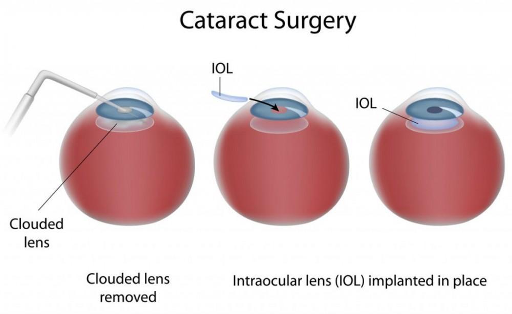
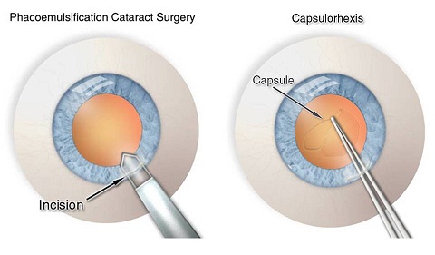
TREATMENT
Cloudy lens needs a Replacement with a clear lens by means of surgery
- LASER CATARACT SURGERY
- MICS IOL
- PHACO IOL –multifocal, monofocal, toric
Phacoemulsification with intra ocular Lens Implantation has been the most accepted and successful treatment for Cataracts worldwide. Cloudy Lens is removed from the eye through a tiny opening and a foldable artificial lens is implanted into the eye.
Contact us today to know cost of cataract surgery in Delhi and Meerut
at. 01141556445,26680397,
GLAUCOMA SERVICES
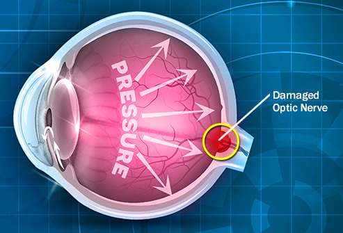
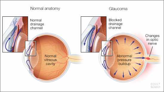
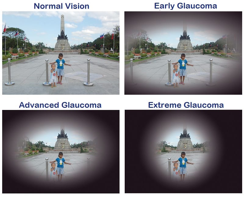
What is glaucoma ?
The increased eye pressure, called intraocular pressure, can damage the optic nerve of the eye , which transmits images to your brain. If the damage continues, glaucomacan lead to permanent vision loss. Without treatment,glaucoma can cause total permanent blindness within a few years.
SYMPTOMS OF GLAUCOMA :
- Severe throbbing eye pain
- Redness of eye
- Headache (on the same side as the affected eye)
- Blurry or foggy vision
- Seeing ” Halos” around lights,
- Fixed Mid-Dilated pupil
- Nausea and Vomiting
INVESTIGATIONS AND TREATMENT MODALITIES :
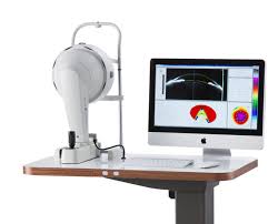
1.PERIMETRY- Humphrey feild analyser
2. OCT
3. YAG LASER
4. UBM
5. SURGERY
Glaucoma is a life-long condition and needs a longer follow-up with your eye doctor.
Contact us today to know cost of glaucoma surgery in Delhi and Meerut
at. 01141556445,26680397,
RETINA & UVEA SERVICES
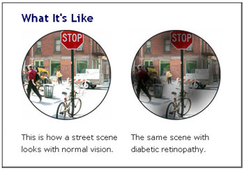
DIABETIC RETINOPATHY
Why eye examination is mandatory for diabetics ?
Diabetes can seriously harm your vision
Diabetic retinopathy is effect of high blood sugar on eyes.
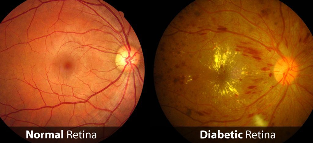
Most diabetics develop some Retinopathy over time
Any diabetic patient might develop retinopathy.
Longer the duration of diabetes , higher will be risk of retinopsthy
SYMPTOMS
You might not have symptoms in the early stages of diabetic retinopathy. As the condition progresses, diabetic retinopathy symptoms may include:
- Spots or dark strings floating in your vision (floaters)
- Blurred vision
- Fluctuating vision
- Impaired color vision
- Dark or empty areas in your vision
- Vision loss
TREATMENT
Treatment, largely depends on the type of diabetic retinopathy you have and how severe it is mainly
Contact us today to know cost of laser treatment for diabetics in Delhi and Meerut
at. 01141556445,26680397,
ARMD
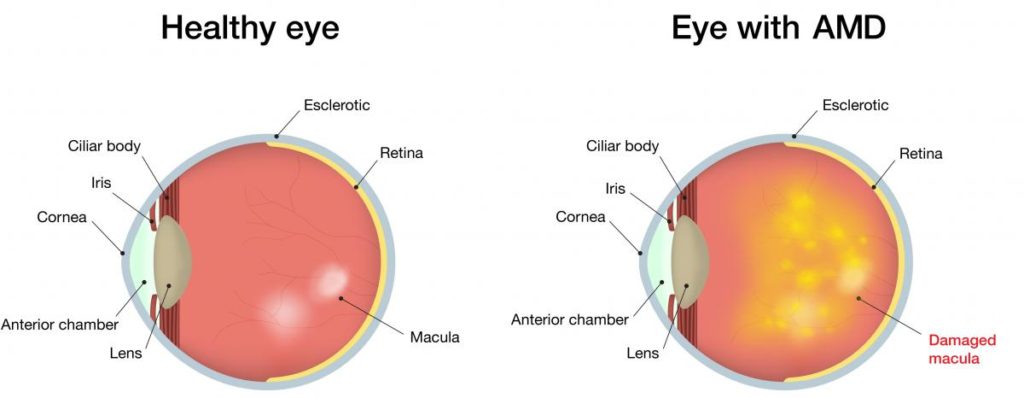
When you do start experiencing vision loss from age-related macular degeneration, symptoms can include:
- Blurred or “fuzzy” vision.
- Straight lines, such as sentences on a page, appearing wavy or distorted.
- Blurry areas on a printed page.
- Difficulty reading or seeing details in low light levels.
- Extra sensitivity to glare.
ARMD TREATMENT
Your Treatment Options
- Anti-VEGF(Accentrix, Lucentis etc.) drugs. Your doctor injects these medications into your eye. …
- Laser therapy. Your doctor may suggest a treatment with high-energy laser light that can sometimes destroy actively growing abnormal blood vessels from AMD.
- Photodynamic laser therapy.Injected drug is activated by laser light.
Symptoms
- The sudden appearance of many floaters — tiny specks that seem to drift through your field of vision.
- Flashes of light in one or both eyes (photopsia)
- Blurred vision.
- Gradually reduced side (peripheral) vision.
- A curtain-like shadow obscuring your field of vision.
Treatment and Drugs
- Retinal Tears Retinal holes or tears will usually need to be treated with laser treatment or cryotherapy (freezing), to seal the retina to the back wall of the eye again. …
- Detached Retina Retinal detachments may require surgery to return the retina to its proper position in the back of the eye.
RETINITIS PIGMENTOSA
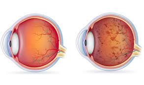
Retinitis Pigmentosa Symptoms
- Loss of night vision. Night blindness is when you cannot see anything in the dark. …
- Gradual loss of peripheral (side) vision. This is known as “tunnel vision.” You may find you bump into things as you move around. …
- Loss of central vision. Some people also have problems with central vision. …
- Problems with color vision.
TREATMENT
There’s no cure for retinitis pigmentosa, but doctors are working hard to find new treatments. Our only goal is to rehabilitate individuals by means of low vision aids.
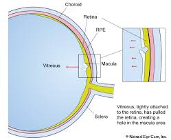
SYMPTOMS OF MACULAR HOLE
Macular holes usually develop over time, so you may not notice any symptoms until your vision is affected. Early signs include blurring and distortion of your vision, and you may notice straight lines (such as window frames, telegraph poles or lines of text) appearing bent or wavy.
TREATMENT
Vitrectomy is the most common treatment for macular holes. In this surgical procedure, the vitreous gel is removed to stop it from pulling on the retina, and most commonly a gas bubble is placed in the eye to gently hold the edges of the macular hole closed until it heals.
MACULAR EDEMA
Macular edema is the build-up of fluid in the macula, an area in the center of the retina. The retina is the light-sensitive tissue at the back of the eye and the macula is the part of the retina responsible for sharp, straight-ahead vision. Fluid buildup causes the macula to swell and thicken, which distorts vision.
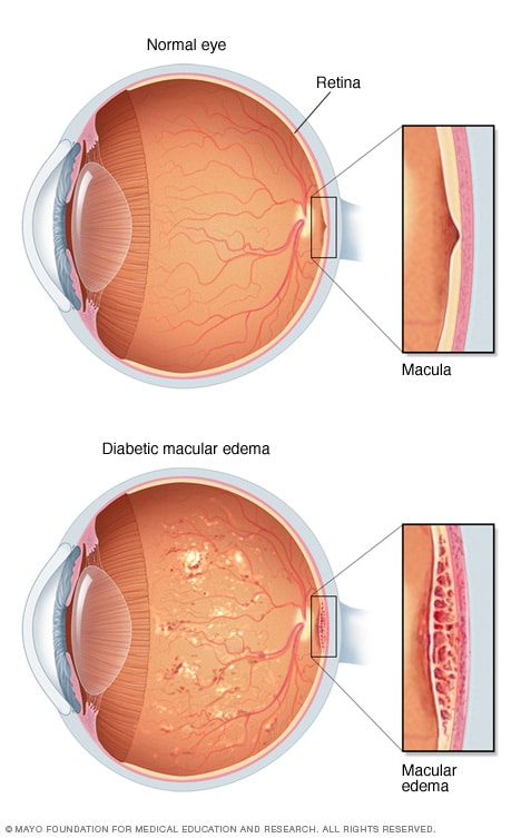
Symptoms
Diabetic macular edema doesn’t always cause symptoms.
But you may:
- Have images directly in front of you appear blurry or wavy
- See colors that seem “washed out”
If this happens to you, see your doctor right away.
Treatment
To treat diabetic macular edema, doctors may use drugs that are injected into your eyes to help stop leaking, and to slow the growth of new blood vessels. These drugs include:
- Accentrix (ranibizumab)
- Eylea (aflibercept)
- Iluvien (fluocinolone acetonide)
- Lucentis (ranibizumab)
- Macugen (pegaptanib)
In severe cases, you may also have laser photocoagulation. A doctor will use a tiny laser on your eye to seal leaking blood vessels. You may need more than one treatment to control the problem. It’s usually not painful, but you may have slight stinging feeling when the laser touches you.
Sometimes steroid injections may help.
Another treatment is a surgery called vitrectomy. This is usually done because of bleeding (not macular edema), and doctors take out the fluid that is clouding your vision and replace it with a clear solution.
RETINAL DETACHMENT
Retinal detachment describes an emergency situation in which a thin layer of tissue (the retina) at the back of the eye pulls away from its normal position.
Retinal detachment separates the retinal cells from the layer of blood vessels that provides oxygen and nourishment. The longer retinal detachment goes untreated, the greater your risk of permanent vision loss in the affected eye.
Warning signs of retinal detachment may include one or all of the following: the sudden appearance of floaters and flashes and reduced vision. Contacting an eye specialist (ophthalmologist) right away can help save your vision.
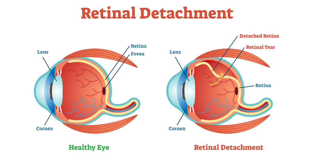
Symptoms
Retinal detachment itself is painless. But warning signs almost always appear before it occurs or has advanced, such as:
- The sudden appearance of many floaters — tiny specks that seem to drift through your field of vision
- Flashes of light in one or both eyes (photopsia)
- Blurred vision
- Gradually reduced side (peripheral) vision
- A curtain-like shadow over your visual field
TREATMENT
- Laser surgery (photocoagulation). The surgeon directs a laser beam into the eye through the pupil. …
- Freezing (cryopexy). After giving you a local anesthetic to numb your eye, the surgeon applies a freezing probe to the outer surface of the eye directly over the tear.
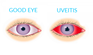
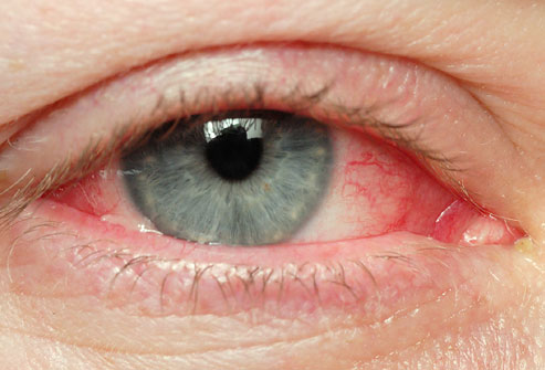
UVEITIS/IRITIS
Uveitis is a form of eye inflammation. It affects the middle layer of tissue in the eye wall (uvea). Uveitis (u-vee-I-tis) warning signs often come on suddenly and get worse quickly. They include eye redness, pain and blurred vision
The signs, symptoms and characteristics of uveitis may include:
SYMPTOMS
- Eye redness.
- Eye pain.
- Light sensitivity.
- Blurred vision.
- Dark, floating spots in your field of vision (floaters)
- Decrease
TREATMENT
- Prescription eye drops in combination with anti-inflammatory medications. …
- Ocular anti-inflammatory injections – injections may be to the outside or inside of the eye. …
- Systemic or oral administration of steroids, other immunosuppressant or anti-metabolite drugs.
ALL TYPES OF UVEITIS ARE TREATED HERE.
CORNEAL SERVICES
CORNEAL ULCER
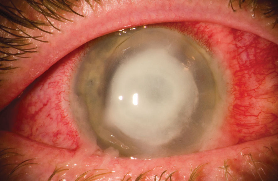
TREATMENT MODALITIES PROVIDED BY US :
CORNEAL SCRAPING ; where a portion of ulcer removed with help of blade locally to find out causitive organism.
CORNEAL DEBRIDEMENT ; where ulcer as a whole is removed topically and investigated further to find out cause.
THERAPEUTIC PENETRATING KERATOPLASTY ;
WANT A CURE FOR NON RESPONDING ULCERS?
Therapeutic Penetrating Keratoplasty is a surgical procedure where diseased cornea is replaced with healthy donor cornea.
Contact us today at Delhi and Meerut
KERATOCONUS
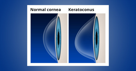
Keratoconus is a condition in which normal shape of cornea becomes conical .
It is generally observed in :
Those having high cylindrical power
Progressive change in spectacle power
Family History positive
Who Are at RISK ?
HOW WE COME TO KNOW ?
HOW DO WE GET RID OF THAT?
CORNEAL COLLAGEN CROSSLINKING PROCEDURE(C3R) :
Procedure where transparent portion cornea is treated with riboflavin drops after removing superficial layer followed by exposure to ultraviolet radiation.
PAINLESS PROCEDURE
Contact us today if anyone in your family member having keratoconus at delhi and Meerut
CONTACT LENSES
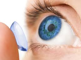
A contact lens is a thin, curved lens placed on the film of tears that covers the surface of your eye. The lens itself is naturally clear, but is often given the slightest tinge of color to make them easier for wearers to handle. Today’s contact lenses are either hard or soft. Most people wear the latter form now, but it wasn’t too long ago that contact lenses were even glass blown!
OCULOPLASTY
: plastic surgery of the eye and adjacent parts (such as the tear ducts or eyelids)
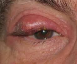
CHALAZION
- A chalazion is a small, slow-growing lump or cyst that develops within the eyelid. They are not usually painful and rarely last longer than a few weeks. A chalazion can develop when a meibomian gland at the edge of an eyelid becomes blocked or inflamed. These glands produce oil that lubricates the surface of the eye.
TREATMENT
- Warm compresses. Soak a clean washcloth in hot water and hold it to your eyelid for 10–15 minutes at a time, 3–5 times a day. …
- Antibiotics. …
- Surgery to drain the area
- Do not squeeze or try to pop a stye or chalazion.